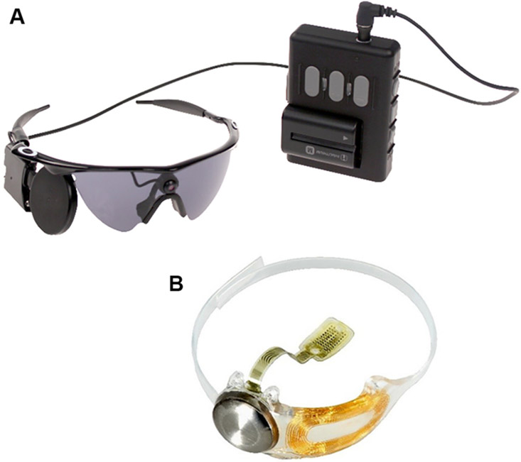
Fig. 3.
(A) The external components; glasses (left) and video processing unit (right) of the Argus II implant. (B) The ocular components of the Argus II implant, including a scleral band, internal coil (for power and data transfer) and the electrode array which is attached to the epiretinal surface with a tack. Images courtesy of Second Sight Medical Products, USA.

