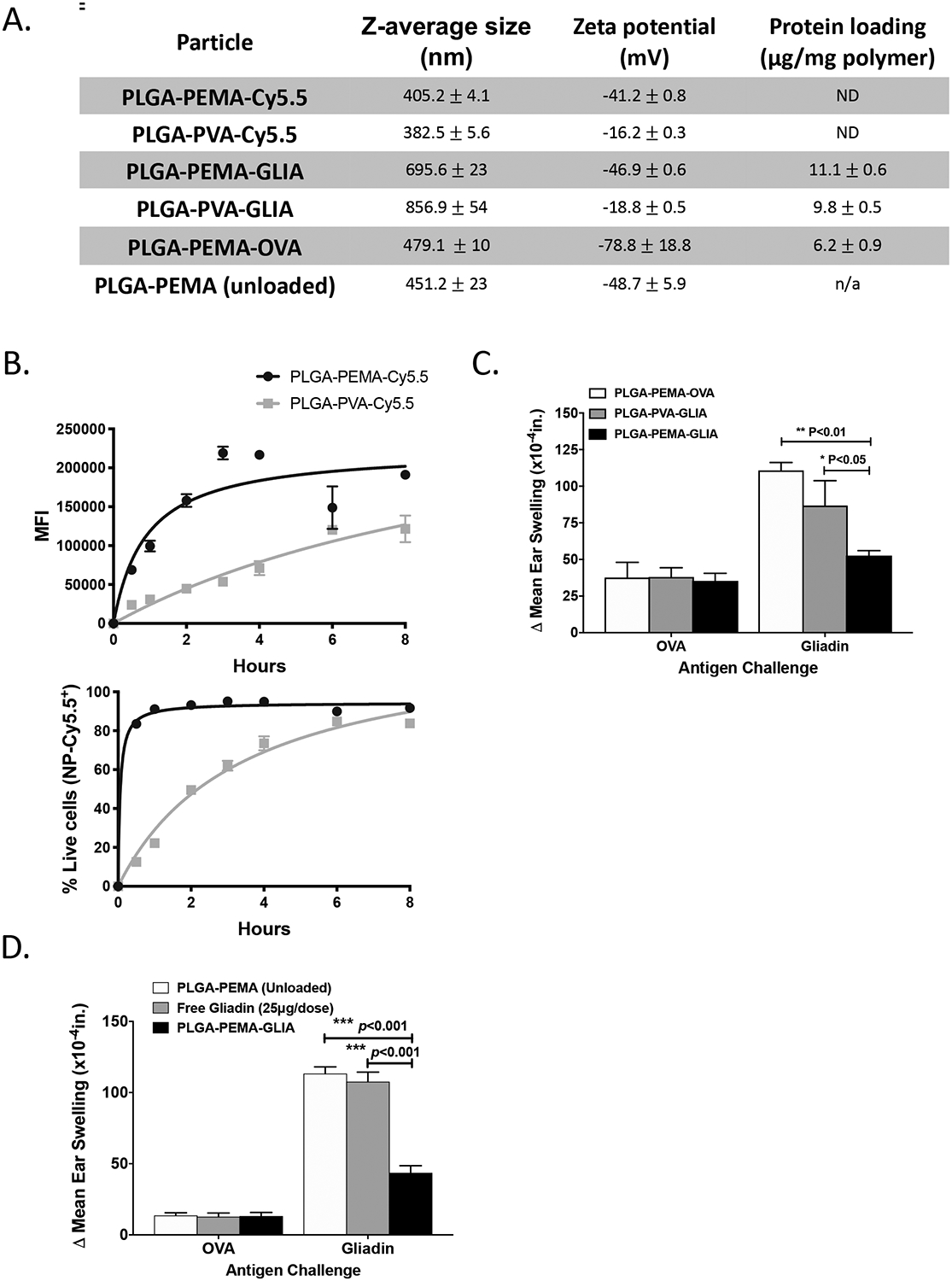
Figure 1: Development of Tolerogenic Immune-Modifying Nanoparticles encapsulating gliadin (TIMP-GLIA) (I).

A) Six different formulations of nanoparticles were prepared for testing, using either PEMA or PVA as stabilizing surfactant, and encapsulating either Cy5.5 dye, gliadin or ovalbumin (or remaining unloaded). Size, charge and protein loading were measured (mean +/− SD). B) PLGA-PEMA-Cy5.5 or PLGA-PVA-Cy5.5 particles were added to bone marrow-derived macrophage cultures. Cells were analyzed by flow cytometry for mean fluorescence intensity, or for percentage of Cy5.5+/DAPI- live cells (triplicates; mean +/− SD). C) Intravenous treatment effect of PLGA-PEMA-GLIA, PLGA-PVA-GLIA or PLGA-PEMA-OVA in the gliadin DTH mouse model. Ear thickness was measured 24h after injection of either gliadin or ovalbumin (n=5; Δ mean ear thickness +/− SEM; x10−4in). D) Treatment effect of PLGA-PEMA-GLIA, soluble gliadin or PLGA-PEMA (unloaded) in the gliadin DTH mouse model (n=5; Δ mean ear thickness +/− SEM; x10−4in). Statistical analyses were performed using one-way ANOVA and Tukey’s multiple comparison test (C, D; *p≤0.05, **p≤0.01, ***p≤0.001)
