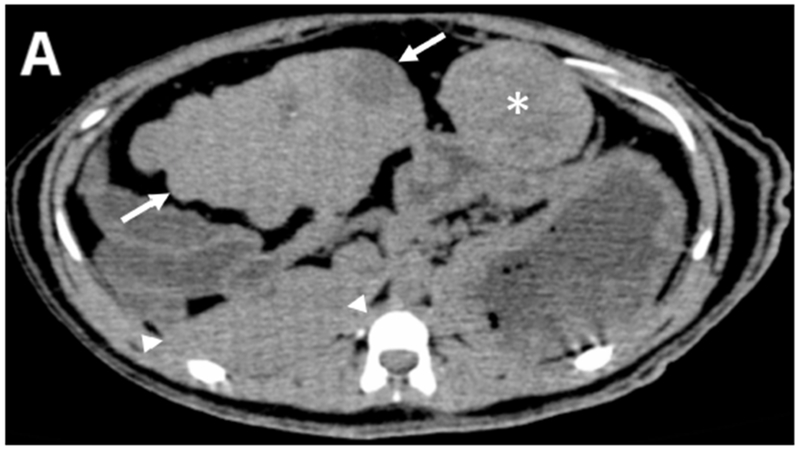
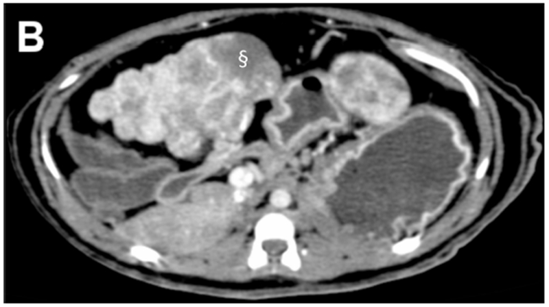
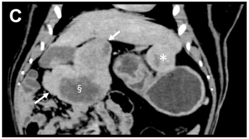
Figure 1.


Pre-procedural contrast enhanced multidetector CT images of two tumors in a woodchuck. (A) Axial image of the upper abdomen without contrast. (B) Early arterial phase image at the same level. (C) Coronal reconstruction of the portal venous phase. The larger, 6 cm pedunculated tumor (arrows) attached to the left medial and quadrate lobes and the smaller 3.2 cm tumor (*) showed early arterial enhancement with washout evident in the smaller tumor. A non-enhancing, low attenuation component of the larger tumor (§) represented an area of necrosis. An area of liver without tumor is shown (arrowheads).
