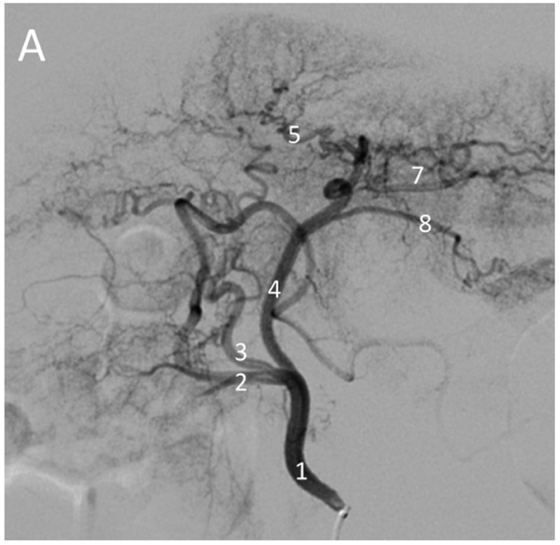
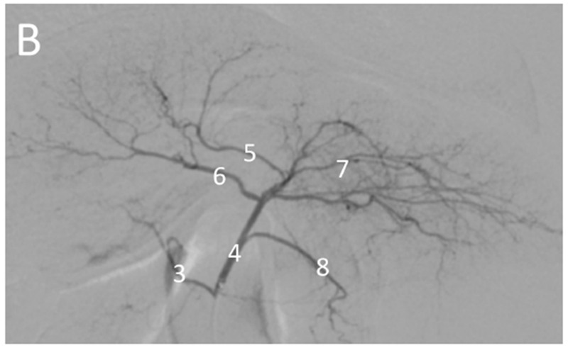
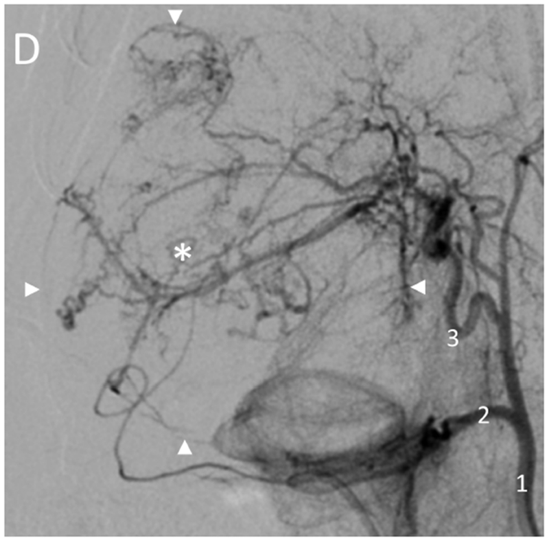
Figure 2.


Hepatic angiography of normal (non-tumor) and tumor-bearing woodchucks. Normal hepatic vasculature (A) following injection of the hepatic artery (1) or (B) injection of the proximal left hepatic artery (4) showing the gastroduodenal artery (2), right hepatic artery (3), left medial lobe artery (5), quadrate lobe artery (6), left lateral lobe arteries (7) and gastroesophageal artery (8). In (A), the gastroduodenal and right hepatic arteries arise from a common branch of the hepatic artery with the right hepatic artery giving rise to the quadrate lobe artery in this anatomic variant. In woodchucks with HCC (C, D), celiac artery injection (C) showing an enlarged right hepatic artery (3, arrow) supplying a large HCC (*). Hepatic artery injection (D) showing an enlarged right hepatic artery (3) supplying a large HCC (*) delineated by the arrowheads. Abnormal tumor vasculature is evident within the tumor.
