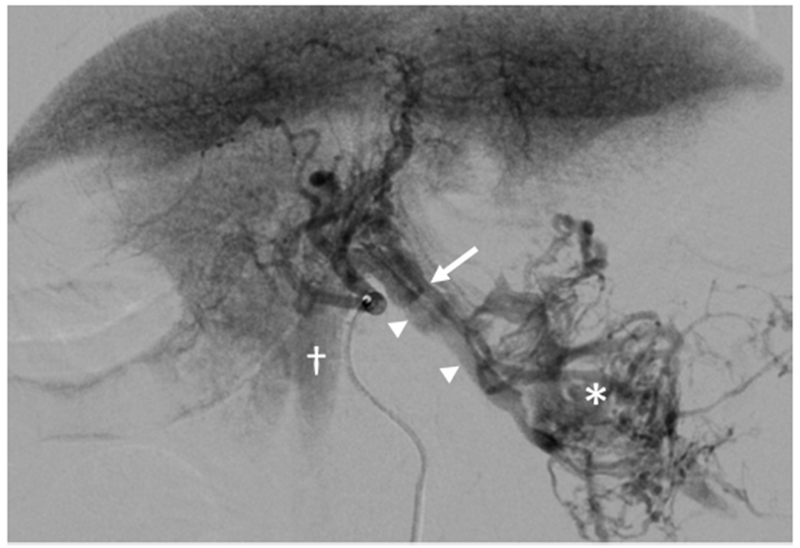
Figure 3.

Large HCC with arteriovenous shunt. The artery (arrow) supplying the HCC (*) is visible with early filling of draining veins (arrowheads) and opacification of the inferior vena cava (†) which contrasts with parenchymal enhancement of normal liver. Arterial and later phase images (Figure E1 A and B) are available online on the article’s Supplemental Material page at www.jvir.org.
