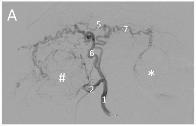
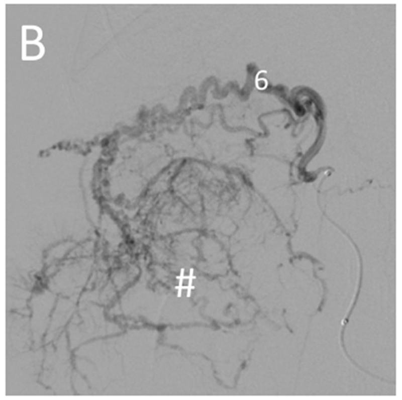
Figure 4.


Selective catheterization of the artery supplying a large tumor. Pre-embolization digital subtraction angiography for the woodchuck shown in Figures 1 and 5. (A) Hepatic artery (1) injection showing the arterial supply of the two tumors: the right tumor (#) by the quadrate lobe artery (6) and the left tumor (*) by the left lateral lobe artery (7). The gastroduodenal artery (2) and left medial lobe artery (5) are also shown. (B) Selective catheterization and angiography of the quadrate lobe artery (6) supplying the tumor on the right (#).
