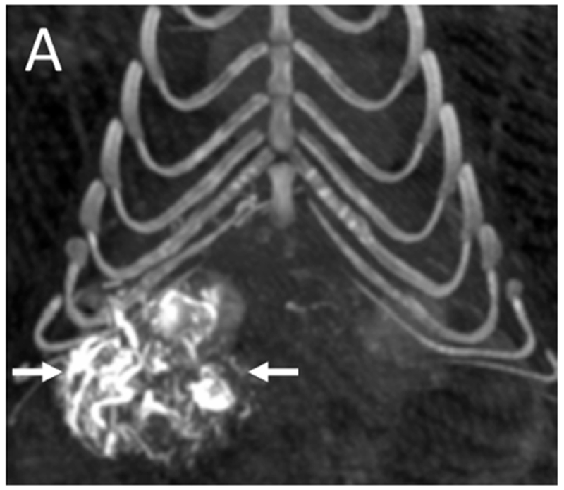
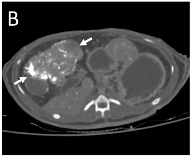
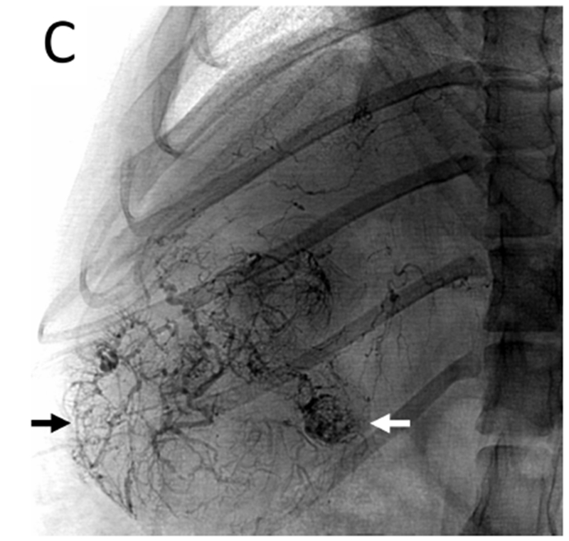
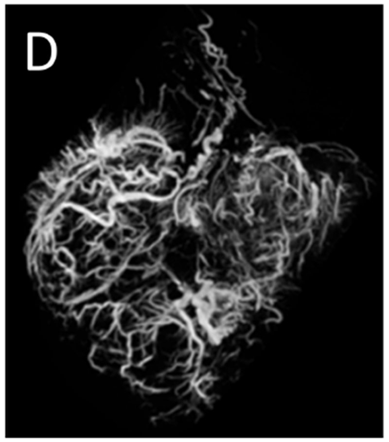
Figure 5.


Post-embolization imaging without contrast administration. Radiopaque drug eluting embolics (arrows) are present within the vasculature of the pedunculated tumor from the same woodchuck shown in Figures 1 and 4. No contrast was administered. (A) Coronal maximum intensity projection image of CBCT; (B) axial MDCT; (C) single fluoroscopic image of the abdomen; and (D) microCT of tumor explant.
