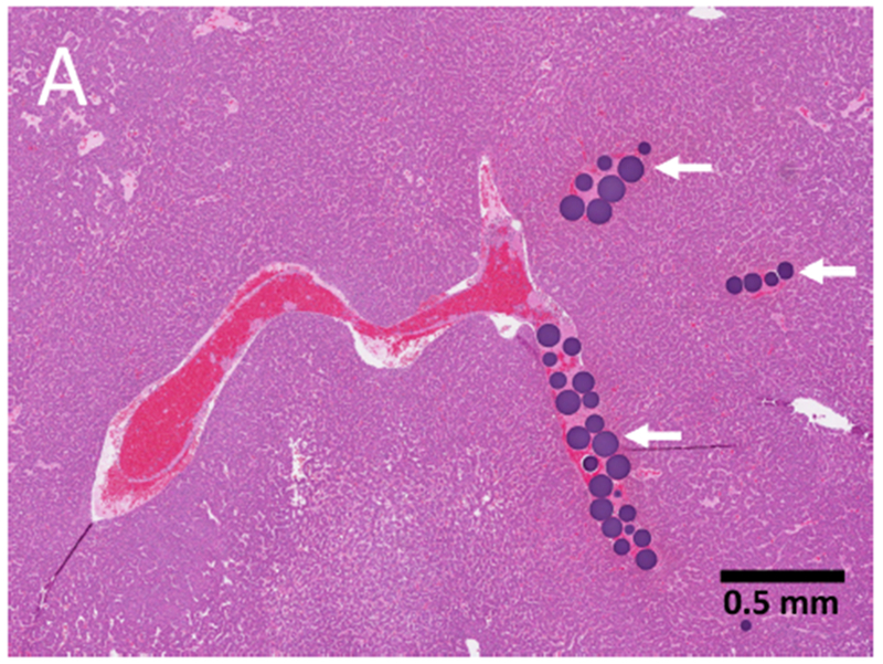
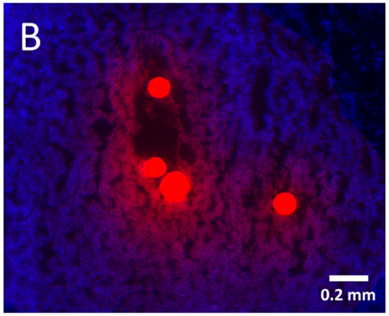
Figure 6.


Hepatocellular carcinoma pathology: drug eluting embolics and drug diffusion into tumor. (A) Histopathology specimen (H&E) of HCC with microspheres (arrows) visible in intratumoral arteries; (B) Fluorescence microscopy of tumor section showing elution of doxorubicin (red) from the microspheres into surrounding tissue (blue, nuclear stain) 45 min post-embolization.
