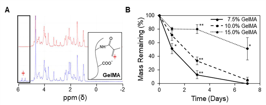
Figure 2: Characterization of the GelMA hydrogel.
(A) Representative 1H NMR spectrums of gelatin (red) and GelMA (blue), with the boxed peaks representing protons attached to the vinyl group of methacrylate (red ╪). (B) Degradation of GelMA hydrogels in collagenase IV shows a slower rate of degradation as GelMA content is increased (n = 3, mean ± STD). One-way ANOVAs with Tukey’s post-hoc comparisons were conducted between groups at each time point. *p < 0.005 and **p < 0.001).

