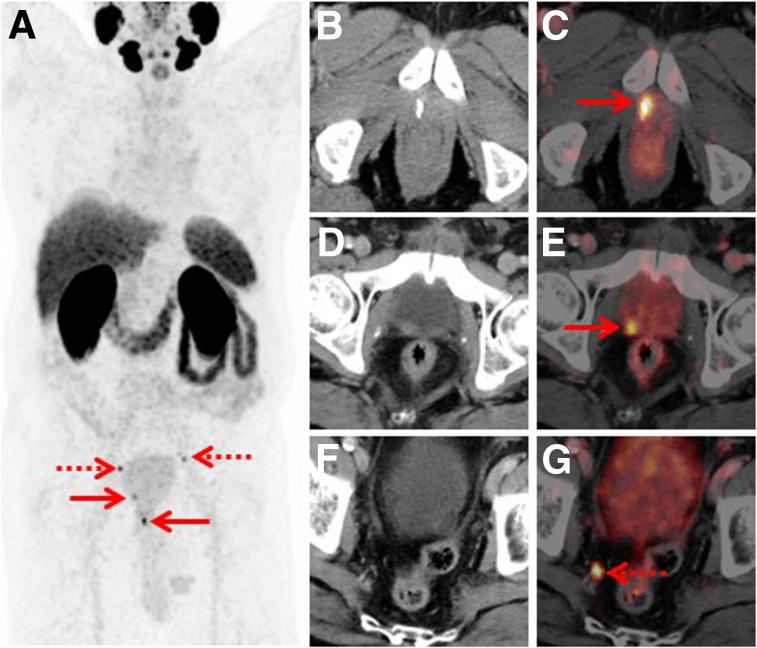
FIGURE 3.
Set of images from 77-y-old patient who underwent radical prostatectomy in 2015 (Gleason score of 9, pT3b, pN1) and was experiencing rising PSA (0.15 ng/mL). (A) Whole-body maximum-intensity projection shows 4 sites with focal PSMA-ligand uptake in pelvis (arrows). (B–G) Axial fused PET/CT and CT images demonstrate local recurrence at anastomosis (B and C, arrow), additional local recurrence at dorsal bladder wall (D and E, arrow), and tiny lymph node metastasis in right pelvis (F and G, arrow). Targeted external-beam radiation treatment lead to subsequent PSA drop.

