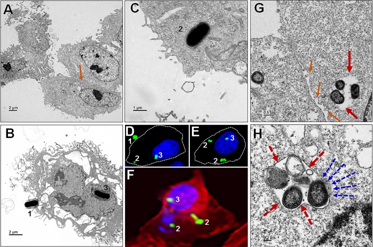
Figure 5.
P. gingivalis manipulation of ARPE cell entry and escape to cytosol. (A) Uninfected control (orange arrow shows the tight junction). (B) Pg interact with ARPE cells (1) and reached the cell nucleus (3). (C) Invaded Pg freely occupy the cytoplasm of ARPE cells (2). (D–F) Confocal images shows the three stages (1. host-pathogen interaction to enter, 2. Invasion and 3, Internalization) and invasion location of the Pg into ARPE cells (membrane, cytoplasm and nucleus), which is consistent to the TEM as shown in (B,C) as well. (D,E) White dotted lines indicates the cell membrane boundary as visualized by F-actin (F). (G,H) The adhesive properties of fimbriae allow P. gingivalis to invade host cells and escape the host immune surveillance. (G) Bacteria in vacuole to evade the host immune defense and survive long (red arrows). Intracellular Pg is found within vesicular structures with single enclosed membranes (dotted red arrows); also noticed Pg organisms inside the retinal epithelial cell, without endocytic vacuoles surrounding them, probably escaped from the vacuole, is evident (H). Also observed loss of integrity of tight junctions in between RPEs (G), compared to uninfected control (A) - orange arrows. Note:- the vacuole membrane breaking and Pg escape to the cytosol (dotted blue arrows). (Scale bar:- A-B: 2 µm, C: 1 µm, G: 0.5 µm, H: 0.2 µm; D, E and F: enlarged region from Figs. 1B and 2B, respectively). Refer the low magnification in Fig. S4 for the complete view.

