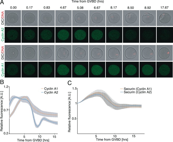
Figure 2.
Oocytes are unable to efficiently target cyclin A1 for degradation. (A) Frames from live cell imaging experiment of oocytes co-injected with cRNAs encoding histone (red) and either cyclin A2 or cyclin A1 (green) fused to fluorescent proteins. Scale bar represents 10 μm. (B) Profiles of fluorescent signal of cyclin A1 (orange, n = 12) and cyclin A2 (blue, n = 7) during meiotic maturation. The curves represent average curve for each construct and the standard deviation error bars are shown. The signal in each cell was normalized to the frame closest to the disassembly of the nuclear membrane (GVBD). The data were obtained from two independent experiments. (C) Profiles of fluorescent signal of securin fused to fluorescent protein in oocytes injected by cyclin A1 (orange, n = 14) or cyclin A2 (blue, n = 10) cRNAs during meiotic maturation. The curves represent average expression curve for all cells in the group and the standard deviation error bars are shown. The signal in each cell was normalized to the frame closest to GVBD. The data were obtained from two independent experiments.

