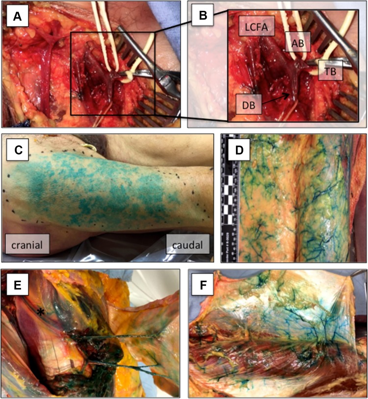
Fig. 1.
Vascular supply of the fascia lata. The lateral circumflex femoral artery (LCFA), the descending (DB), ascending (AB), and transverse branches (TB) were identified first (a, b). Thereafter, the DB was cannulated and 40 ml methylene blue were injected (not shown). After we assessed the maximal length and width of stained skin paddles (c), we elevated the adipocutaneous tissue suprafascially from medial to lateral (d). To protect the peri-fascial blood supply, we preserved some adherent adipose tissue. The fascia lata was supplied by septocutaneous (e) or musculocutaneous (f) perforators. Asterisk (*) marks the DB of the LCFA within the intermuscular septum after retraction of the rectus femoris muscle

