
FIGURE 4.
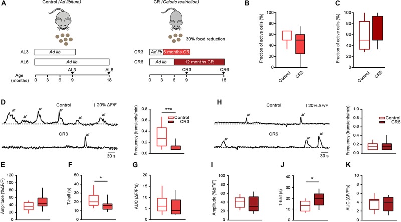
Effect of caloric restriction on spontaneous Ca2 + signals in middle-aged and old mice. (A) Schematic description of caloric restriction protocols used in these experiments. In the control group (left), the animals had unlimited access to food (ad libitum). In the CR group (right), mice were daily fed with the 70% of their respective initial ad libitum intake. CR mice were split into two groups based on the starting date and the duration of the CR diet: CR3 mice were under CR from 3 till 9 months of age; CR6 mice were under CR from 6 till 18 months of age. For all CR mice there was no return to ad libitum feeding before the end of experiment. (B) Box-and-whisker plot illustrating the fractions (per mouse) of spontaneously active microglia in 9–11 months old control (n = 10 mice, 34 cells) vs. CR3 (n = 7 mice, 26 cells) mice. (C) Box-and-whisker plot illustrating the fractions (per mouse) of spontaneously active microglia in 18–21 months old control (n = 14 mice, 46 cells) vs. CR6 (n = 8 mice, 26 cells) mice. (D,H) Left: representative spontaneous microglial Ca2 + transients (arrows) measured in control (top) and CR (bottom) mice. Right: box-and-whisker plots illustrating the median (per cell) frequency of spontaneous Ca2 + transients in microglia from the respective groups. (E–K) Box-and-whisker plots illustrating the median (per cell) amplitude (E,I), T-half (F,J) and AUC (G,K) of the spontaneous Ca2 + transients in control vs. CR mice (n = 19 cells (control), 12 cells (CR3) for 9–11 months old mice; n = 28 cells (control), 16 cells (CR6) for 18–21 months old mice). Statistical differences were determined using Mann-Whitney test (*p < 0.05, ***p < 0.001).