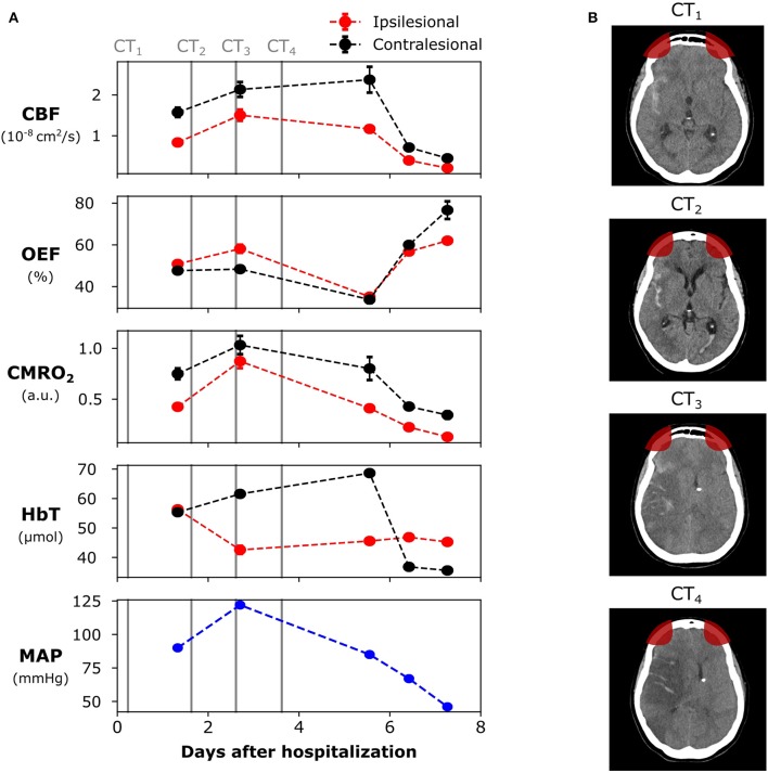
Figure 1.
Evolution of the brain lesion in a 62 years old female patient following a high-grade aneurysmal subarachnoid hemorrhage (aSAH). (A) Neurophysiological parameters measured with the diffuse optical system, as well as the systemic mean arterial pressure (MAP). (B) Computed tomography (CT) images at different days during hospitalization (marked as vertical lines in the left panel). The patient died 9 days after hospitalization. The red areas in the CT images represent the optical sensitivity region. The error bars of each point represent the standard deviation of each parameter across the monitoring time-window. For some days, the standard deviation was too small to be shown. CBF, cerebral blood flow; OEF, oxygen extraction fraction; CMRO2, cerebral metabolic rate of oxygen; HbT, total hemoglobin concentration.

