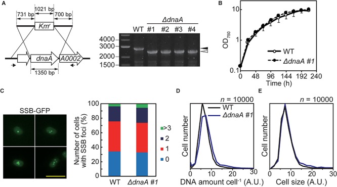
FIGURE 4.

Effect of deleting dnaA on the growth and DNA replication of Synechococcus sp. PCC 7002. (A) Chromosomal dnaA was replaced with the gene encoding kanamycin resistance (Kmr) through homologous recombination. Insertion of Kmr into the chromosomal dnaA locus was confirmed using PCR with the primers indicated by the arrows. The wild type (WT) was used as a negative control. The black and white arrowheads indicate the bands amplified from the WT and mutated chromosomes, respectively. The four independent colonies of the dnaA-deficient mutant were analyzed. (B) Growth of WT and ΔdnaA clone #1 in an inorganic medium with illumination (70 μmol m–2 s–1). Other ΔdnaA clones (#2–#4) are shown in Supplementary Figure S6B. (C) Frequencies of cells exhibiting zero (blue), one (red), two (deep blue), or >3 (green) SSB-GFP foci in WT and ΔdnaA clone #1 cultures 12 h after inoculation (n > 300 cells, each strain). For reference, a representative fluorescence image of WT expressing SSB-GFP (green fluorescence) is shown (one to four SSB-GFP foci are shown). Scale bar = 5 μm. The construction of SSB-GFP expresser is described in Supplementary Figure S3. (D,E) Flow cytometric analysis showing the distributions of the DNA level per cell (D) and the cell volumes (E) of cultures of the WT (black line) and ΔdnaA clone #1 (blue line) 12 h after inoculation, respectively. DNA was stained with SYTOX Green, and the levels were determined according to the intensity of the green fluorescence.
