Figure 4.
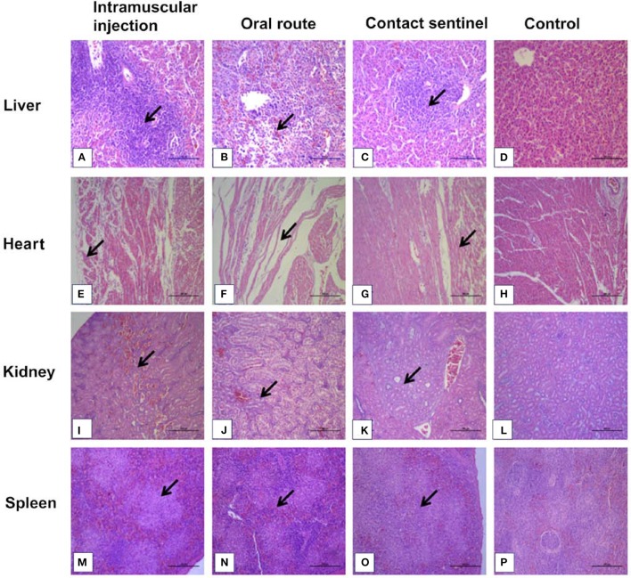
Histopathology in liver, heart, kidney, and spleen from dead chickens at 5 to 7 dpi (stained with HE) inoculated with CH/HBTF/1710. Massive pathological damage was observed in the virus-inoculated chickens. Degeneration, vacuolar necrosis, and basophilic inclusion bodies were present in hepatic cells (A–C). Rupture and necrosis of myocardial fibers were observed in heart tissue (E–G). Swelling and degeneration of renal tubular epithelial cells were present in the kidneys (I–K). Severe reduction and necrosis of lymphocytes were seen in the spleen (M–O). There was no significant histopathological damage present in the liver (D), heart (H), kidney (L), and spleen (P) of chickens from the control group. Scale bar = 200 μm in (A,B), and 100 μm in the other panels. Solid arrows point to lesion areas.

