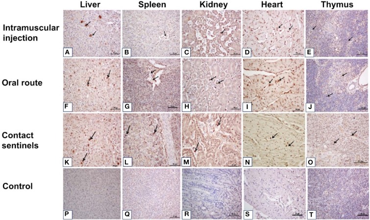
Figure 5.
Immunohistochemical detection of FAdV antigens in the liver, spleen, kidney, heart, and thymus tissues after infection with FAdV CH/HBTF/1710 isolate at 5 to 7 dpi. Positive signals for FAdV antigen were extensively detected by immunohistochemical staining in the liver (A,F,K), spleen (B,G,L), kidney (C,H,M), heart (D,I,N), and thymus (E,J,O) in the two infection groups and the contact sentinels at 5-7 dpi. No FAdV antigen was detected in the liver (P), spleen (Q), kidney (R), heart (S), and thymus (I) in the control group. Solid arrows indicate positive signals for viral antigen. Scale bar = 50 μm.

