Figure 2.

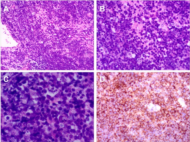
Histopathology of Case 1. (A) Haematoxylin and eosin (H&E) ×10. (B) H&E ×25. (C) H&E ×100. H&E staining for brain tissue biopsy showed diffuse proliferation of large atypical lymphoid cells. (D) Immunohistochemical staining for CD20 showed that most cells were positive.
