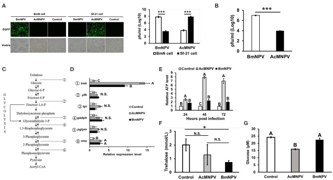
Figure 1.
Virus-host tropisms and host glycolytic activities upon AcMNPV and BmNPV infection. (A) Virus titers were determined by fluorescence intensity and by qPCR analysis at 48 hpi in BmN and Sf-21 cells infected with AcMNPV or BmNPV. (B) Virus titers quantification by qPCR in AcMNPV- or BmNPV infected B. mori larvae at 48 hpi (C) Summary of glycolytic and citrate cycle enzymes in insects. (D) RT-qPCR analysis of glycolytic genes trehalase-2 (treh), phosphofructokinase (pfk), triose phosphate isomerase (tpi), glyceraldehyde 3-phosphate dehydrogenase (gapdh), phosphoglyceromutase (pglym), and enolase (eno) in BmN cells at 48 h after infection with AcMNPV or BmNPV. All of the results were normalized to expression of the 18 S rRNA gene and non-infected control (ΔΔCt). (E) ATP levels were measured at 48 hpi in AcMNPV- or BmNPV-infected cells. All of the results were normalized to those in the non-infected control. Hemolymph trehalose (F) and glucose (G) levels in B. mori larvae were measured at 48 hpi; control larvae were injected with 1X PBS. All the values are the mean ± SEM of three (A,C,G) or four (E,F) replicates. Significances of D, E, F, and G were determined by one-way ANOVA with Tukey's HSD post-hoc analysis; different letters for the treatment group indicate significant differences at P < 0.05. Student's t-test was used for the analysis of A, B, F, *P < 0.05, ***P < 0.001.

