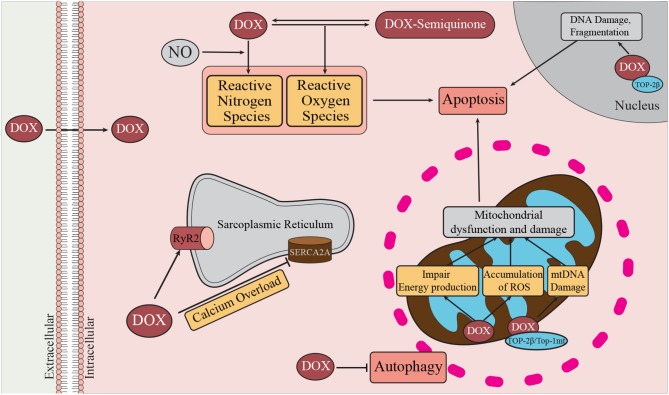
Figure 2.
Mechanisms of DOX-induced cardiotoxicity. DOX enters the cell by passive diffusion, and within the cytoplasm undergoes redox cycling resulting in the generation of ROS and RNS (47). Oxidative and nitrosative stress are known to contribute to activation of cell death pathways, such as autophagy, necrosis, and apoptosis. DOX can promote Ca2+ overload by inhibiting SERCA2a and transiently enhancing the activity of RyR2. DOX can bind cardiolipin, a major inner mitochondrial membrane lipid. At the mitochondria DOX promotes damage to mitochondrial DNA (mtDNA) through interactions with Top-2β, and Top-1mt. In the nucleus, DOX interacts with Top-2β and DNA to form the Top-2β–DOX—DNA cleavage complex (48). By binding to Top-2β, DOX disrupts the rejoining of DNA leading to the accumulation of double stranded DNA breaks thereby triggering apoptosis (49). Damage to mitochondrial DNA affects mitochondrial biogenesis and contributes to mitochondrial dysfunction and therefore reduced ATP production (50).

