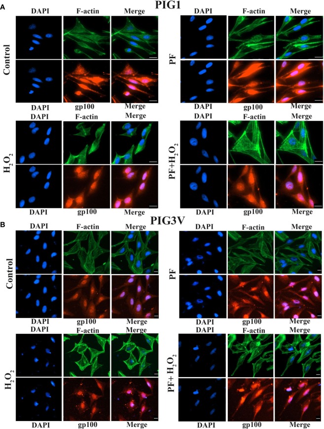
Figure 5.
PF promotes dendrite formation and melanosome transport under H2O2-induced oxidative damage. Melanocytes were exposed to PF for 24 h and were further treated with H2O2 for another 24 h. The dendrite lengths were increased and contained more melanosomes in both stressed PIG1 (A) and PIG3V (B) when pre-treated with PF compared to H2O2 treated only. Then cells were fixed and stained with antibodies recognizing F-actin (phalloidin, green) and melanosome (gp100, red). Nuclei were counterstained with DAPI (blue). Immunofluorescence was used to determine the localization and expression of melanosomes and F-actin in the cells. Scalebar = 20µm. H2O2, hydrogen peroxide; PF, Paeoniflorin.

