Fig. 7.

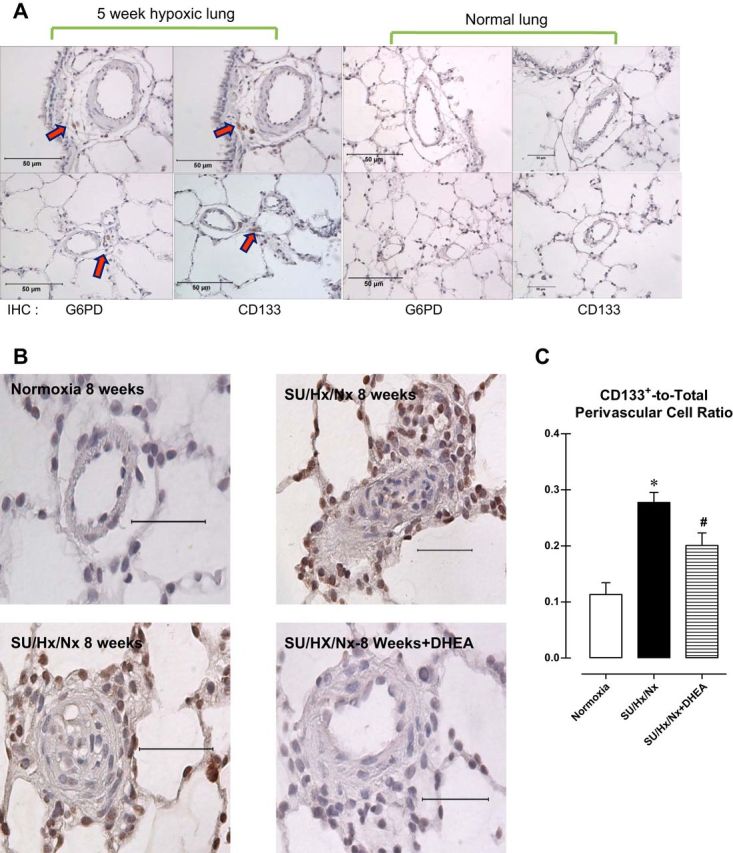
A: immunohistochemical staining of sections of rat lung exposed to normoxia or hypoxia for 5 wk. Arrows indicate cells positive for G6PD and CD133+. B: immunohistochemical staining for CD133 in pulmonary arteries from a normotensive rat (top left), pulmonary arterial hypertensive rat (SU/Hx/Nx 8 wk: top right and bottom left), and pulmonary arterial hypertensive rat treated with DHEA (bottom right). C: summary data showing CD133+ cell accumulation in perivascular regions was decreased by DHEA treatment.
