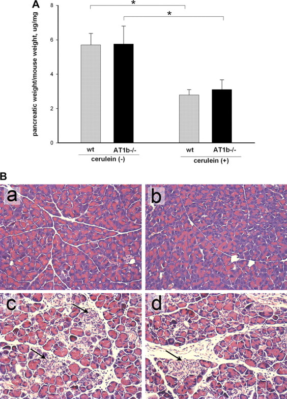
Fig. 6.

Pancreatic weight and hematoxylin and eosin stains in AT1b −/− mice after repetitive episodes of acute pancreatitis (as described in Fig. 1). A: loss of pancreatic weight relative to total body weight in cerulein-treated mice suggests significant atrophy (8 mice per group), *P < 0.001. B: histological changes in the pancreas of AT1b −/− and WT mice after repetitive episodes of acute pancreatitis (representative picture, hematoxylin and eosin stain, original magnification, ×400). a and b: pancreas from WT (a) and AT1b −/− mice (b) after control saline treatment showed no abnormalities in untreated AT1b −/− mice. c and d: pancreas from cerulein treated WT (c) and cerulein-treated AT1b−/− (d) mice showed severe parenchymal atrophy, dedifferentiation to tubular complexes, and interstitial inflammation. There were no differences in these parameters of chronic pancreatitis between WT and AT1b −/− mice.
