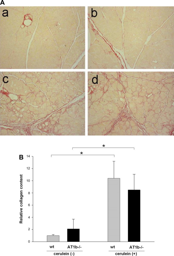
Fig. 7.

Collagen content in the pancreas of WT and AT1b −/− mice after repetitive episodes of acute pancreatitis. A: representative Sirius red staining of pancreatic sections of control pancreas from WT (a) and AT1b−/− (b) mice and from cerulein-treated WT (c) and AT1b−/− (d) mice. Collagen staining appears red (original magnification ×200). B: relative amount of pancreatic collagen quantified by morphometric analysis. Similar to AT1a −/− mice, the absence of AT1b did not alter the accumulation of collagen. Results are expressed as means ± SE, n = 6. *P < 0.01.
