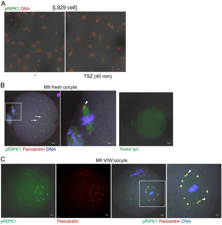
Fig. 5.
Localization of pRIPK1 in vitrified-warmed oocytes. a Specificity of anti-pRIPK1 antibody was confirmed in L929 cells treated with necroptosis-inducing reagents. -, no treatment; TSZ, a mixture of TNFα, LCL161, Z-VAD-FMK. Scale bar represents 20 μm. b Immunofluorescence staining of pRIPK1 in MII oocytes from young mice. The primary antibodies used are, anti-pRIPK1 (1:150, green) and anti-pericentrin (1:500, red). DNA was counterstained with TOPRO-3-iodide (1:250). Overlapped signals of pRIPK1 and pericentrin are visualized in yellow. Arrows indicate pericentrin-positive MTOCs and arrowhead indicates the spindle pole. The white boxed area is enlarged on the right. White scale bar represents 20 μm or 10 μm in the enlarged image. c A representative image showing co-localization of pRIPK1 and pericentrin. The white boxed area is enlarged four times. The primary antibodies used are, anti-pRIPK1 (1:150, green) and anti-pericentrin (1:500, red). Arrowheads indicate the spindle poles. The white boxed area is enlarged on the right. White scale bar represents 10 μm or 5 μm in the enlarged image

