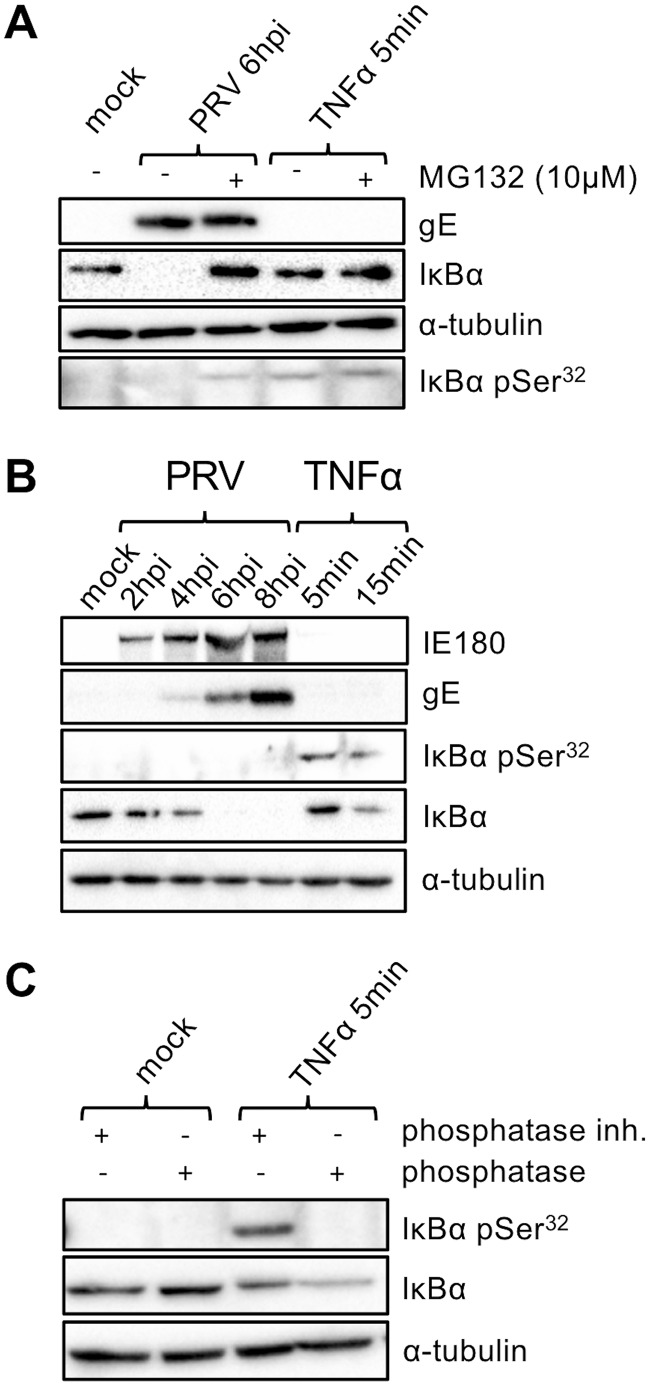
FIG 5.
(A) Western blot analysis of total IκBα and IκBα phosphorylated at serine 32 (pSer32) either in PRV Kaplan-infected ST cells (6 hpi; MOI, 10 PFU/cell) or in TNF-α-treated ST cells (5 min posttreatment, 100 ng/ml) in the presence or absence of the proteasome inhibitor MG132 (10 μM; which was added for 4 h starting at 2 hpi in PRV-infected cells and which was used for preincubation for 4 h in TNF-α-stimulated cells). (B) Western blot analysis of total IκBα and IκBα phosphorylated at serine 32 either in PRV Kaplan-infected ST cells (0, 2, 4, 6, and 8 hpi; MOI, 10 PFU/cell) or in TNF-α-treated ST cells (5 and 15 min posttreatment, 100 ng/ml). (C) Western blot analysis of total IκBα and IκBα phosphorylated at serine 32 in mock-treated or TNF-α-treated ST cells (5 min, 100 ng/ml) incubated or not with bacteriophage lambda protein phosphatase. The Western blots shown are representative examples from three independent repeats of the experiments.

