FIG 7.
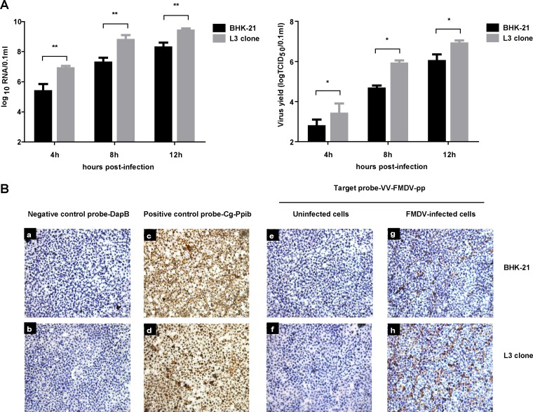
Knockout of hnRNP L in cells promoted FMDV viral RNA synthesis. (A) BHK-21 cells or L3 clones were infected with FMDV at an MOI of 1. The resulting viruses were harvested at 4 h, 8 h, and 12 h and the supernatant was analyzed for viral RNA and virus titer. (B) Seed cells of BHK-21 cells or L3 clone cells in growth medium on chamber slides were infected with FMDV at an MOI of 1. At 8 h postinfection, the cells were used for the RNAscope assay. Target probes were hybridized for 2 h at 40°C, followed by a series of signal amplification and washing steps. Hybridization signals were detected by chromogenic reactions using DAB chromogen followed by 1:1 (vol/vol)-diluted hematoxylin counterstaining. Only in vitro samples with an average of at least 1 positive (brown) dot per cell were included for analysis. Slides were examined and captured for each section using Leica Application Suite (LAS) v3.8 (Leica Microsystems).

