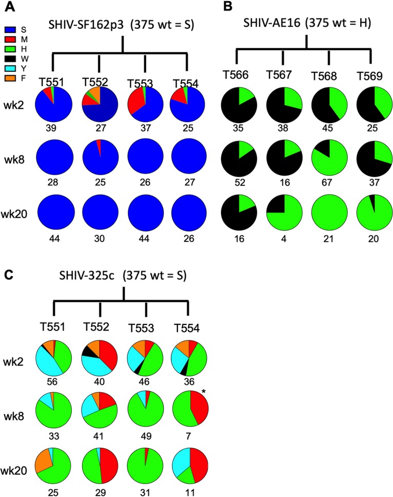
FIG 3.

Sequence analysis of SHIV evolution in infected rhesus monkeys. Single-genome amplification was performed with plasma viral RNA on weeks 2, 8, and 20 for animals infected with SHIV-SF162p3 (A), SHIV-AE16 (B), and SHIV-325c (C) pool variants. Numbers under the circle plots indicate total sequence value per sample. Note that week 4 was assessed instead of week 8 for T562 due to low viral loads at week 8.
