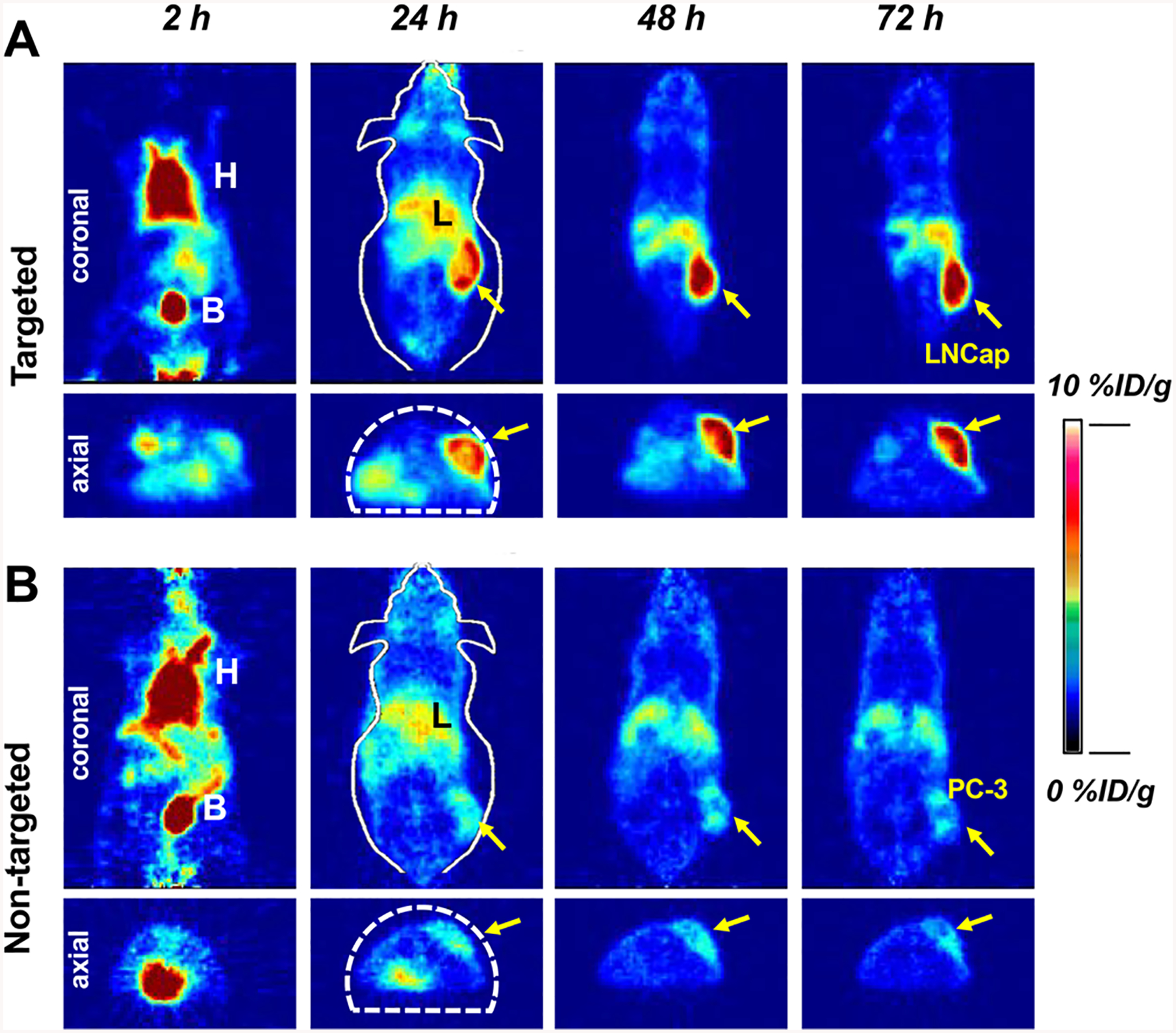
Figure 3.

In vivo cancer-targeted PET imaging of 89Zr-DFO-PSMAi-PEG-Cy5-C′ dots intravenously injected into (A) LNCap, targeted group, and (B) PC-3, nontargeted group, tumor-bearing mice. H: heart; B: bladder; L: liver. Both LNCap and PC-3 tumors were marked with yellow arrows.
