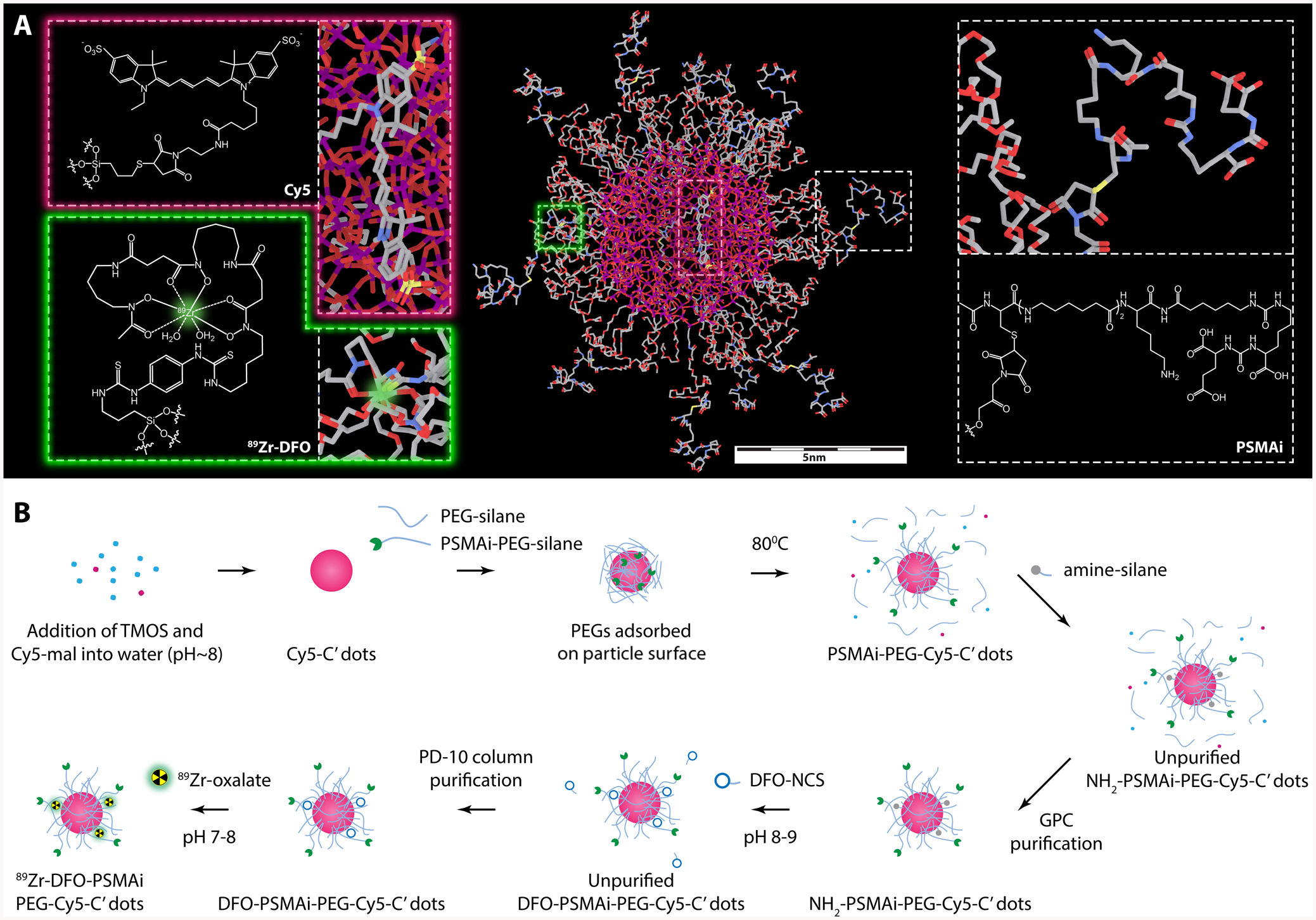
Scheme 1. (A) Three-Dimensional (3D) Rendering of Core-Shell Structured 89Zr-DFO-PSMAi-PEG-Cy5-C′ Dot Prostate Cancer-Specific Targeting Probe;a (B) Schematic Illustration Showing the Synthesis of 89Zr-DFO-PSMAi-PEG-Cy5-C′ Dotsb.

aThe core of the nanoparticle is a Cy5 dye-encapsulating silica scaffold with its physical size controlled to be around 3–4 nm. The shell includes a protective layer of short PEG chains (MW: ~500 g/mol), radionuclide chelators, DFO, for 89Zr labeling, and PSMAi peptides for active PCa targeting. bCy5-silane was reacted with TMOS in DI water at room temperature with the pH adjusted to ~8 to form Cy5-C′ dots. Then PEGylation and peptide-functionalization steps were introduced to form PSMAi-PEG-Cy5-C′ dots. As-synthesized PSMAi-PEG-Cy5-C′ dots were then surface-functionalized with amine groups by reacting with amine-silanes to form NH2-PSMAi-PEG-Cy5-C′ dots. The GPC-purified NH2-PSMAi-PEG-Cy5-C′ dots were mixed with DFO-NCS to form DFO-PSMAi-PEG-Cy5-C′ dots. Finally, 89Zr was attached to form 89Zr-DFO-PSMAi-PEG-Cy5-C′ dots.
