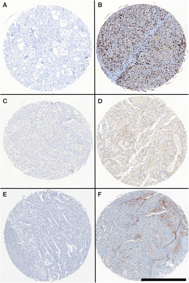
Figure 1.

CD8-, PD-1-, PD-L1 specific staining in gastric and esophageal adenocarcinoma TMA. Representative CD8, PD-1, and PD-L1-specific staining in TMA punches. Specimens were stained with CD8- (B), PD-1- (D), and PD-L1- (F) specific reagents. (A, C, E) refer to punches stained with isotype control reagents. Scale bar: 500 μm.
