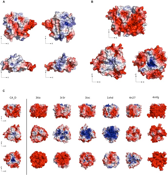
FIGURE 2.
Electrostatic surface potential of CA_D. (A) CA_D Monomer and (B) CA_D Trimer electrostatic surface potential color-coded from red (negative potential) to blue (positive potential). (C) The surface potential of CA_D compared to mesophilic γ-CA homologs (Escherichia coli, 3tio; Salmonella enterica, 3r3r; Anaplasma phagocytophilum, 3ixc; Bacillus cereus, 1xhd; Brucella abortus, 4n27; Clostridium difficile, 4mfg). Unit: –5 to +5 kbT/e (kb as the Boltzmann constant, T as the temperature in Kelvin and e as the charge of an electron).

