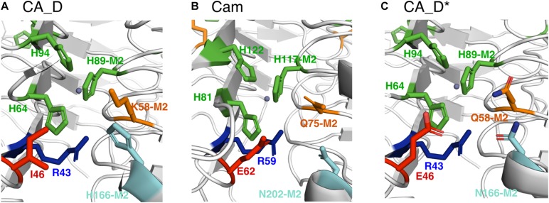
FIGURE 4.
Comparison of selected active site residues. (A) CA_D (crystal structure), (B) Cam (crystal structure) from M. thermophila (PDB ID: 1qrg), and (C) CA_D* including selected mutations such as I46E, K58Q, H166N (M2: indicates that residue belongs to the adjacent monomer). The central zinc ion is depicted as a gray sphere.

