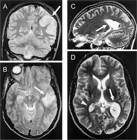
Figure 1.

A and B, Single, large, tumorlike lesion of the brain (arrows) associated with cerebral edema in a 23-year-old patient with postinfluenzal encephalopathy noted on T2-weighted MRIs. Adenovirus DNA was detected in a CSF sample obtained from this patient 1 day earlier. C and D, MRIs for this patient 2 months later that reveal regression of this single lesion (arrow) but also an additional lesion (arrowhead).
