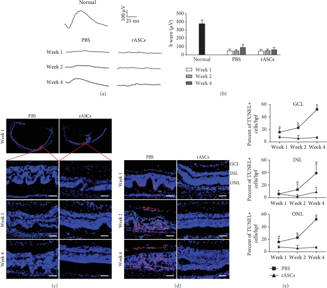
Figure 2.

Protective effects of rASCs on retina in SI-induced rat RDD model. 3 × 10⁵ rASCs were transplanted into the vitreous chamber. (a) Representative ERG waveforms recorded at different time points (the calibration indicates 100 μV vertically and 25 ms horizontally). (b) Quantitative analysis of ERG b-wave amplitude (n = 10). (c) Representative DAPI-stained micrographs of retinal samples (scale bar = 50 μm). (d) Representative micrographs of retinal cryosections stained with TUNEL (scale bar = 50 μm). (e) Statistical analysis of the percentage of the apoptotic cells in GCL, INL, and ONL (n = 12). Results are expressed as mean ± SEM; ∗P < 0.05 compared with the rASC group. GCL: ganglion cell layer; INL: inner nuclear layer; ONL: outer nuclear layer.
