
Figure 6.
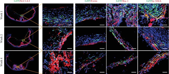
Representative immunofluorescence images of ERM-like structures by grafted GFP-labeled ASCs in the vitreous chamber from different time points. The cellular compartments of ERM-like structures include Müller cells (SLC1A3+), RPE cells (Ezrin+), microglial cells (Iba-1+), and myofibroblast cells (α-SMA+); arrows point to the GFP+/α-SMA+ cells (scale bar = 50 μm). ERM: epiretinal membrane; RPE cells: retinal pigment epithelial cells.