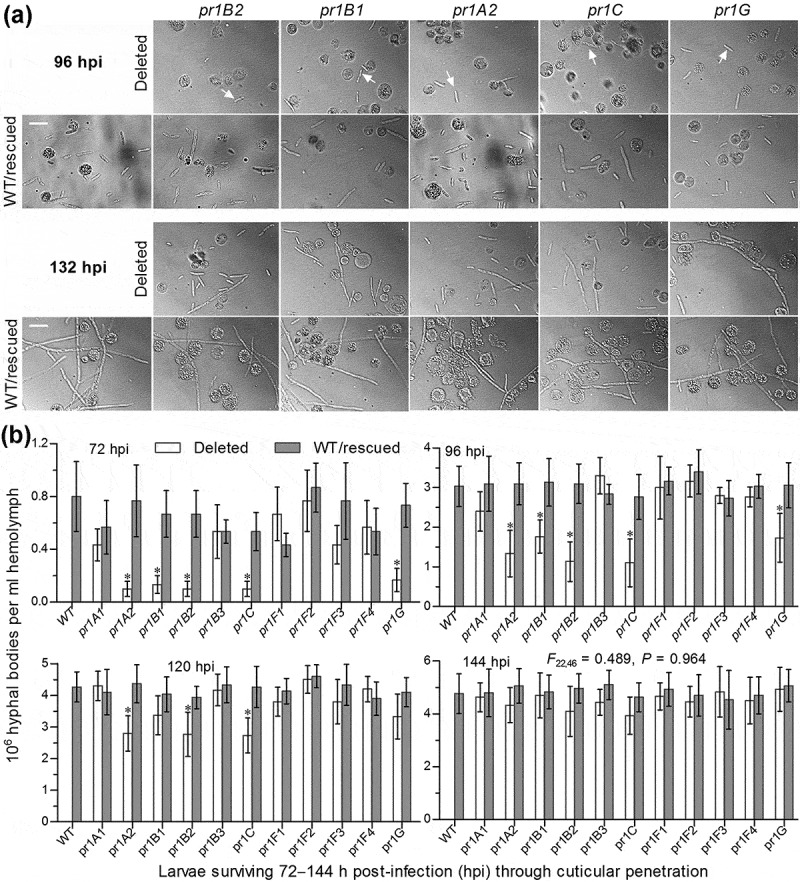
Figure 5.

Impacts of each pr1 deletion on development of hyphal bodies in the hemolymph of G. mellonella larvae after topical application (immersion) of a 107 conidia/ml suspension for normal cuticle infection. (a) Microscopic images (scale = 20 μm) for abundance of hyphal bodies (arrowed) in the hemolymph samples taken from the larvae surviving 96 and 132 h post-infection (hpi). Spherical and subspherical cells are insect hemocytes. (b) Concentrations of hyphal bodies quantified from the hemolymph samples taken from the larvae surviving 72–144 hpi. The asterisked Δpr1 means differ significantly from those of the corresponding control strains unmarked (Tukey’s HSD, P < 0.05). Error bars: SD from three replicates.
