Figure 2.
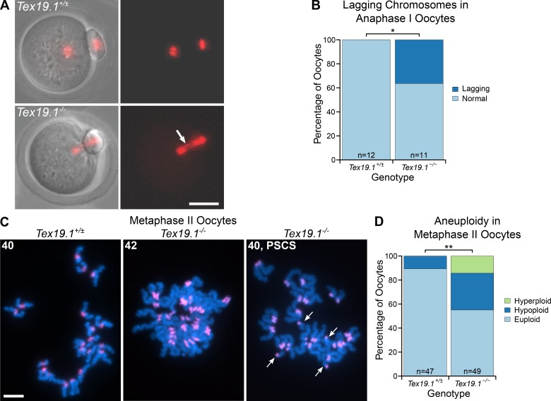
Tex19.1−/− oocytes missegregate homologous chromosomes and prematurely separate sister chromatids during meiosis I. (A) Live imaging of meiosis I in Tex19.1+/± and Tex19.1−/− oocytes. Chromatin was visualized with histone H2B-RFP (red). Lagging chromosomes are indicated with an arrow. Scale bar, 50 µm. (B) 36% of Tex19.1−/− anaphase I oocytes (n = 12) but no Tex19.1+/± anaphase I oocytes (n = 11) contained lagging chromosomes (*, Fisher’s exact test, P < 0.05). Data are from six Tex19.1+/± and three Tex19.1−/− females. (C) Chromosome spreads from metaphase II oocytes. DNA was visualized with DAPI (cyan) and centromeres by major satellite FISH (red). The number of chromatids is indicated. An aneuploid Tex19.1−/− oocyte with 42 chromatids but no overt premature sister chromatid separation (PSCS) and a euploid Tex19.1−/− oocyte with 40 chromatids and PSCS (arrows) are shown. Scale bar, 10 µm. (D) 31% of metaphase II Tex19.1−/− oocytes were hypoploid and 14% hyperploid compared with 11% hypoploid and 0% hyperploid for metaphase II Tex19.1+/± oocytes (**, Fisher’s exact test, P < 0.01; n = 47, 49). Data are from 8 Tex19.1+/± and 12 Tex19.1−/− females.

