Figure 4.
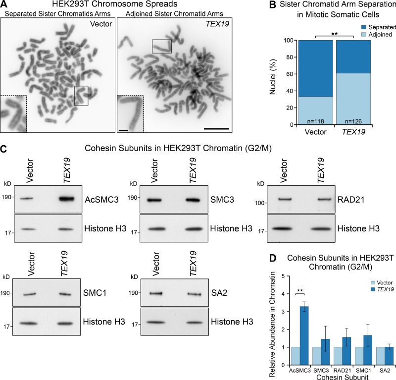
Ectopic expression of TEX19 promotes sister chromatid cohesion in mitotic somatic cells. (A and B) Photographs (A) and quantification (B) of sister chromatid separation in HEK293T cells. Chromosome spreads from HEK293T cells transfected with human TEX19 or empty vector were classed as having separated sister chromatids if most chromosomes had a visible gap between chromosome arms. Scale bar, 10 µm; inset scale bar, 2 µm. Quantification of four independent experiments; n indicates the total number of chromosome spreads. 33% (n = 126) of metaphase spreads from cells transfected with TEX19 had separated sister chromatids compared with 67% (n = 118) of controls (**, Fisher’s exact test, P < 0.01). (C and D) Representative Western blots (C) and quantification (D) of cohesin subunits in chromatin from HEK293T cells transfected with either human TEX19 or empty vector, synchronized to enrich for cells in G2/M. Cohesin abundance was normalized to histone H3 and quantified relative to empty vector transfections. Means ± SD are indicated. Expression of TEX19 induces a 3.3-fold increase in chromatin-associated AcSMC3 (**, t test, P < 0.01; n = 3). Each pair of histone H3 and cohesin bands are from the same gel lane; the high concentration of histones in chromatin causes the sample to spread laterally at low molecular weights (Fig. S5 C).

