Figure 7.
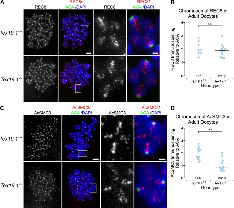
Tex19.1−/− oocytes have reduced levels of chromatin-associated AcSMC3 cohesin. (A and C) Tex19.1+/± and Tex19.1−/− prometaphase I chromosome spreads immunostained with anti-centromere antibodies (ACA; green), DAPI (blue), and either anti-REC8 (A, red) or anti-AcSMC3 (C, red) antibodies to visualize cohesin subunits. Example individual bivalents (boxes) are magnified and shown in the righthand panels. Single-channel images of anti-AcSMC3 and anti-REC8 are also shown in grayscale. Scale bars 10 µm; inset scale bars 2 µm. (B and D) Quantification of anti-REC8 (B) and anti-AcSMC3 (D) immunostaining in prometaphase I oocyte chromosomes. Individual bivalents were distinguished by DAPI staining, and total cohesin immunostaining on each bivalent was measured relative to ACA. The median of the ratios for each oocyte is plotted; horizontal lines indicate median ratios for each genotype. REC8 immunostaining is not significantly different in Tex19.1−/− oocytes (ns, Mann-Whitney U test, not significantly different; n = 8, 12), but AcSMC3 is significantly reduced to 45% of the level detected in Tex19.1+/± controls (**, Mann-Whitney U test, P < 0.01; n = 10, 10). Data are from seven Tex19.1+/± and five Tex19.1−/− females for REC8 and from four Tex19.1+/± and four Tex19.1−/− females for AcSMC3.

