Figure S2.
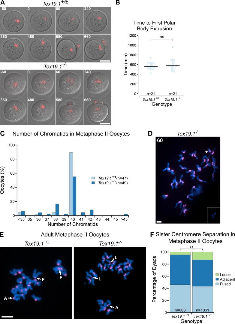
Meiotic chromosome segregation in adult Tex19.1−/− oocytes. (A) Live imaging of meiosis I in Tex19.1+/± and Tex19.1−/− oocytes. Chromatin was visualized with histone H2B-RFP (red). Time relative to GVBD in minutes is indicated in the top left corner of each image. Data are derived from six Tex19.1+/± and three Tex19.1−/− females across seven microinjection and imaging sessions. Scale bar, 50 µm. (B) Beeswarm plot showing the time to polar body extrusion relative to GVBD in Tex19.1+/± and Tex19.1−/− oocytes. Median values are indicated with a horizontal line (ns, Mann-Whitney U test, no significant difference; n = 21, 21). (C) Histogram showing the percentage of Tex19.1+/± and Tex19.1−/− metaphase II oocytes analyzed in Fig. 2 (C and D) that contained the indicated number of chromatids. Note the presence of aneuploid oocytes with odd numbers of chromatids, indicating potential premature segregation of sister chromatids. Of the seven hyperploid Tex19.1−/− oocytes, two exhibited cytologically detectable premature separation of sister chromatids; four had ≥21 dyads exhibiting intact sister chromatid cohesion, indicating missegregation of homologues had occurred; and one hyperploid Tex19.1−/− oocyte exhibited both these traits. (D) Chromosome spreads from an extreme hyperploid Tex19.1−/− metaphase II oocyte containing 60 chromatids. This oocyte contains >20 dyads indicating that homologous chromosomes missegregated without undergoing premature separation of sister centromeres during meiosis I in this oocyte. Note that a pair of broken chromatids lacking centromeric FISH signals (inset) was located just outside the field of view in this spread. These could potentially represent sister chromatids that broke away from their centromeres during preparation of the chromosome spreads, possibly from the centromere pair indicated with an asterisk. Scale bar, 10 µm. (E) Chromosome spreads from Tex19.1+/± and Tex19.1−/− metaphase II oocytes. DNA was visualized with DAPI (cyan), and centromeres were detected by FISH for major satellites (red). Sister centromere separation was scored as fused (F, one continuous centromeric domain between two chromatids), adjacent (A, two separate distinct centromeric domains separated by less than half the width of the chromatid arm), or loose (L, two separate distinct centromeric domains separated by more than half the width of the chromatid arm). Examples of these configurations are annotated. Scale bar, 10 µm. (F) Percentage of metaphase II dyads with different levels of sister centromere separation. 46% of dyads in 863 Tex19.1+/± oocytes have a fused configuration, 49% adjacent, and 5% loose. 43% of dyads in 1061 Tex19.1−/− oocytes have a fused configuration, 44% adjacent, and 13% loose (**, Fisher’s exact test, P < 0.01). Data are derived from 8 Tex19.1+/± and 12 Tex19.1−/− females.

