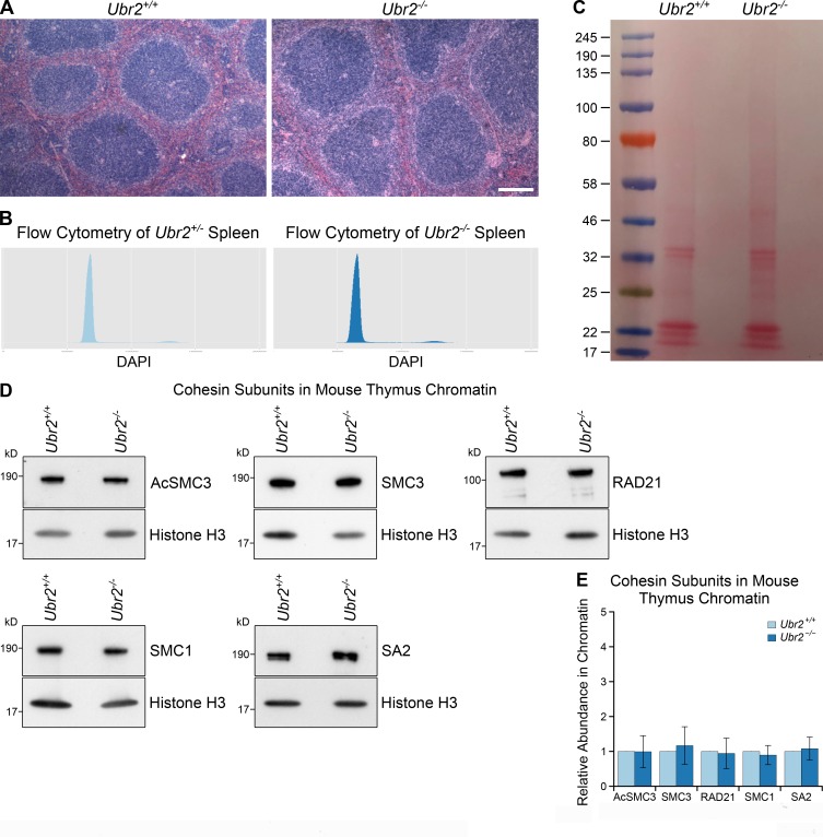
Figure S5.
Additional data from Ubr2−/− mice. (A) Hematoxylin and eosin–stained paraffin sections of Ubr2+/+ and Ubr2−/− spleen. Loss of Ubr2 does not dramatically alter the histological appearance or cell type composition of the spleen. Scale bar, 100 µm. (B) Flow cytometry of Ubr2+/− and Ubr2−/− spleens. Loss of Ubr2 does not grossly perturb the cell cycle distribution in this tissue. (C) Ponceau S stained membrane of chromatin preparations from Ubr2+/+ and Ubr2−/− spleen before Western blotting. Chromatin preparations have high concentrations of histone proteins, which cause the gel lanes to broaden in the low molecular weight (15–25-kD) region. This phenomenon means that some bands visualized with anti-histone H3 antibodies have different widths from bands visualized with antibodies against higher molecular weight cohesins (e.g., anti-SMC3, anti-acetylated SMC3) in this study, despite originating from the same membrane. (D and E) Representative Western blots (D) and quantification (E) from three pairs of Ubr2+/+ and Ubr2−/− mice for the abundance of cohesin subunits (AcSMC3, SMC3, RAD21, SMC1, SA2) in thymus chromatin. Histone H3 was used as a loading control. Cohesin abundance was normalized to histone H3 and quantified relative to Ubr2+/+ mice (graph indicates mean ± SD). The abundance of cohesin subunits in the thymus of Ubr2−/− mice was not significantly different from Ubr2+/+ controls (t test; n = 4).

