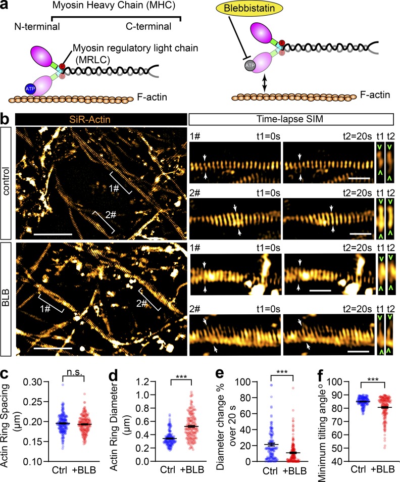
Figure 4.
Short-term inactivation of NM-II affects the diameter and angle of axonal actin rings, but not their periodic spacing. (a) Cartoon showing the organization of actomyosin structure in nonmuscle cells, with the ATP-binding site in the head domain of MHC annotated. Blebbistatin blocks NM-II ATPase activity leading to its detachment from F-actin. (b) In cultured hippocampal neurons, endogenous periodic axonal actin rings were labeled using SiR-actin and live imaged using 2D SIM. Representative time-lapse SIM images of axonal actin rings are shown before (control) and after short-term blebbistatin treatment (10 µM, 30–60 min). Bracketed regions are magnified in right panels. Dynamic diameter changes of actin rings are annotated with arrowheads. Scale bars = 5 µm (left) and 1 µm (right). (c–f) Quantification of the spacing (c), diameter (d), fluctuation of actin ring diameter (e), and minimum tilting angles (f) of the periodic actin rings along the axon. Data represent mean ± SEM, n = 167 (control) and 186 (blebbistatin [+BLB]), representing numbers of axonal actin rings analyzed. Values were measured from three independent cultures (***, P < 0.001, two-tailed unpaired t test). n.s., not significant.

