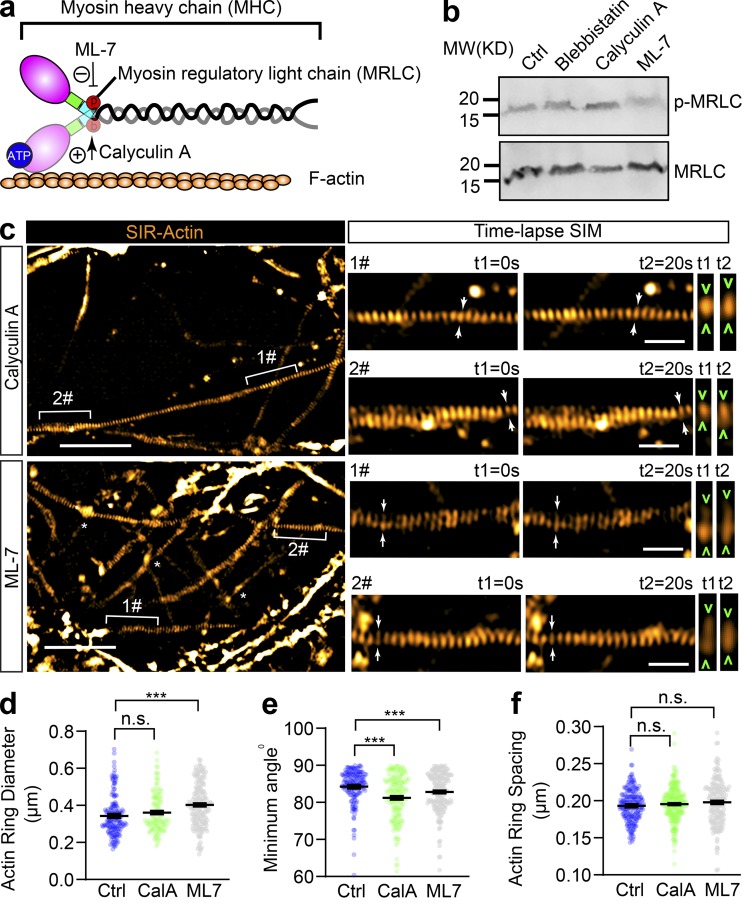
Figure 5.
Inhibition of MRLC phosphorylation slightly affects the diameter and tilting angle of axonal actin rings, but not their periodic spacing. (a) Cartoon showing the organization of actomyosin structure in nonmuscle cells, with the effecting sites of ML-7 and Calyculin A on MRLC annotated. (b) Western blot showing the level of diphosphorylated MRLC (p-MRLC) following 30 min treatment with blebbistatin (10 µM), ML-7 (10 µM), and Calyculin A (50 nM). (c) In cultured hippocampal neurons, endogenous periodic axonal actin rings were labeled using SiR-actin and live imaged using 2D SIM. Representative time-lapse SIM images of axonal actin rings are shown following 30 min of treatment with ML-7 (10 µM) or Calyculin A (50 nM), respectively. Bracketed regions are magnified in right panels. Dynamic diameter changes of actin rings are annotated with arrowheads. Scale bars = 5 µm (left) and 1 µm (right). (d–f) Quantification of the diameter (d), minimum tilting angle (e), and spacing (f) of the periodic actin rings along the axon. Data represent mean ± SEM; n = 167 (control [Ctrl]), 170 (Calyculin A [CalA]), and 175 (ML-7), values are labeled on the panels, representing numbers of axonal actin rings analyzed. Values were measured from three independent cultures (***, P < 0.001, two-tailed unpaired t test). MW, molecular weight; n.s., not significant.

