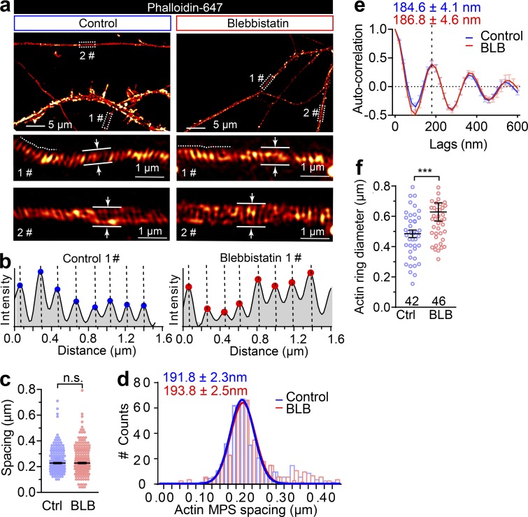
Figure S3.
Short-term inactivation of NM-II increases the axon diameter without affecting the actin ring periodicity. (a) DIV14 rat axons were stained for endogenous F-actin (phalloidin) and imaged with 3D SIM; two different boxed regions magnified with maximum z-projections shown in the lower panels. Axon diameters were measured as the average of a 1-µm segment. (b) Periodic actin peaks were identified using the find peak function of BAR collection in ImageJ (simple moving average = 1), along the line profiles as shown with dashed lines in 1# of panel a. x values of the peaks were extracted (and the distance between adjacent peaks is shown in Fig. 5 f) in both untreated (control) and short-term (60 min) blebbistatin-treated axons. (c and d) Comparison of spot plot (c) and Gaussian fitting curve (d) of periodic actin spacing distribution in control and blebbistatin-treated neurons. Data represent mean ± SEM; n = 300 (control) and 316 (blebbistatin treated) for periodicity quantification. (e) Autocorrelation analysis of the actin periodicity of control and blebbistatin-treated axons. Data represent mean ± SEM; n = 10 (control) and 8 (blebbistatin-treated) axon segments were measured. (f) Quantification of actin diameters in control and blebbistatin-treated axons. Data represent mean ± SEM; n = 42 (control) and 46 (blebbistatin-treated) actin rings diameters were measured. Values were measured from three independent cultures (**, P < 0.05; ***, P < 0.001, two-tailed unpaired t test). n.s., not significant.

