Figure S4.
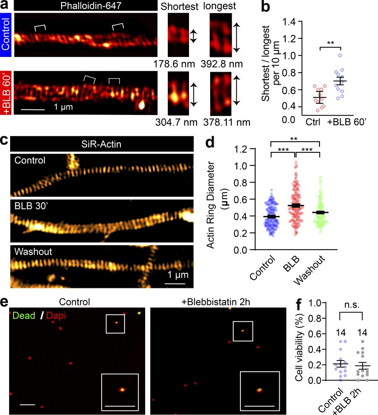
The effect of blebbistatin on periodic axon actin rings is reversible. (a) SIM images of endogenous F-actin (phalloidin) along the axon of a DIV14 rat hippocampal neuron before and after short-term blebbistatin treatment (10 µM, 60 min). Bracketed regions are magnified on the right, and the diameters of actin rings are shown below. (b) Quantification of actin ring diameter fluctuations; the diameters per 10-µm axon segments were measured and quantified. Data represent mean ± SEM; n = 11 (control) and 11 (blebbistatin-treated) axon segments were analyzed. (c) In cultured hippocampal neurons, endogenous periodic axonal actin rings were labeled using SiR-actin and live imaged using SIM. Representative SIM images of axonal actin rings are shown of neurons following DMSO treatment (control), 30-min blebbistatin treatment (BLB), and 12-h incubation after blebbistatin treatment and washout, respectively. Scale bar = 1 µm. (d) Quantification of diameters of the periodic actin rings along the axon. (e) Viability of neurons treated with 10 µM blebbistatin for 120 min. Boxed regions are amplified in the insets. Scale bars = 50 µm. (f) Quantification of viability rate. Data represent mean ± SEM; n values are labeled on the panels, representing numbers of axonal actin rings analyzed. Values were measured from axons of at least three independent cultures (**, P < 0.01; ***, P < 0.001, two-tailed unpaired t test). n.s., not significant.

