Figure 9.
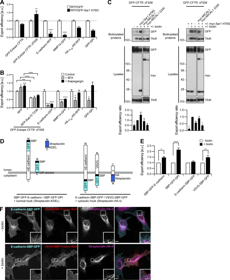
Export of neosynthesized CFTR, CFTR ΔF508, E-cadherin, and MMP14 is sensitive to ER stress. (A) Export efficiency of GFP–Extope CFTR or GFP–Extope CFTR ΔF508 in 293T cells, of E-cadherin–RFP, HA–α2B-AR–RFP, or GFP-GPI in HeLa cells and of MMP14-GFP in HT 1080 cells. n = 3–6. Sar1 H79G increased export of GFP–Extope CFTR ΔF508 but inhibited export of E-cadherin–RFP and MMP14-GFP. (B) Export efficiency of GFP–Extope CFTR ΔF508 in 293T cells and of E-cadherin–GFP, MMP14-GFP, HA–α2B-AR–RFP, or GFP-GPI in HeLa cells. For GFP–Extope CFTR ΔF508, cells were left untreated, incubated with BFA (1.25 µg/ml, 12 h), or thapsigargin (1 µM, 2 h), the latter followed by a 2-h chase (Gee et al., 2011). Cells were collected 36 h after transfection. HeLa cells were incubated with BFA (1.25 µg/ml, 6 h) or thapsigargin (1 µM, 6 h) and collected 12 h after transfection. Basal and ER stress-induced export of GFP–Extope CFTR ΔF508 was inhibited by RFP–Rab10 T23N. ER stress inhibited export of E-cadherin–GFP and MMP14-GFP. n = 4–6. (A and B) The dashed line represents the export efficiency under control conditions. (C) Biotinylation of surface proteins in transfected 293T cells. Export efficiency of GFP–CFTR ΔF508 was quantified using single-transfected cells (left) or cells cotransfected with myc–Sar1 H79G (right) as control. n = 3–5. The Transferrin receptor (TfnR) was used as loading control. In each case, the two parts of the blots are from the same membrane and correspond to identical exposure times. (D) Schematic representation of the RUSH proteins used. (E and F) Transfected HeLa cells were left untreated or incubated with biotin (40 µM, 1 h). (E) Export efficiency of RUSH proteins. n = 3–5. (A, B, C, and E) Data are means ± SEM; *, P < 0.05; **, P < 0.01; ***, P < 0.001; and ****, P < 0.0001. (F) Confocal images after staining with DyLight 650 α-HA. In untreated cells, VSVG-SBP–Fusion Red was in the ER where it colocalized with the Streptavidin-HA-Ii retention hook; E-cadherin–SBP-GFP did not accumulate in the ER and was already found at the plasma membrane. Upon biotin addition, VSVG-SBP-GFP was released from the ER and translocated to the plasma membrane. Insets show magnifications of the boxed areas. Scale bars: 10 µm; scale bars of insets: 4 µm.

