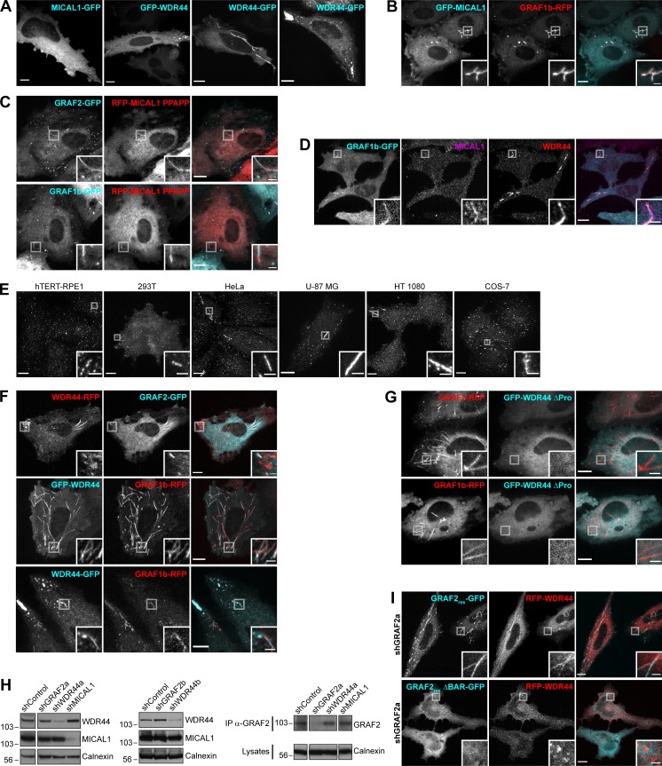
Figure S3.
GRAF1b/2 colocalize with MICAL1 and WDR44. (A) Confocal stacks of transfected HeLa cells showing a similar distribution of N- and C-terminal–tagged MICAL1 and WDR44 (compare with Fig. 2 G). In some cells, WDR44 was found on irregular peripheral patches. (B) Confocal images of transfected HeLa cells showing GRAF1b-RFP on the same intracellular tubules as GFP-MICAL1. (C) Confocal images of transfected HeLa cells showing that although MICAL1 PPAPP did not efficiently colocalize with GRAF1b/2 (Fig. 2 I), it was sometimes found on GRAF-positive tubules (boxed areas). (D) Confocal images of transfected HeLa cells stained with α-MICAL1 and α-WDR44. Endogenous WDR44 and to a lesser extent endogenous MICAL1 colocalized with GRAF1b-GFP. (E) Confocal stacks of hTERT-RPE1, 293T, HeLa, U-87 MG, HT 1080, and COS-7 cells stained with α-WDR44. Endogenous WDR44 tubules of various lengths were found in all but hTERT-RPE1 cells. Tubules were more abundant and longer in HeLa, HT 1080, and U-87 MG cells. (F) Confocal images of transfected HeLa cells showing colocalization of GRAF1b/2 with WDR44 tubules but not with its peripheral patches. (G) Confocal images of transfected HeLa cells showing that WDR44 ΔPro was not recruited to GRAF1b/2 tubules. (H) Western blot analysis of shRNA-transfected HeLa cell lysates. Calnexin was used as loading control. Left and middle: Specific knockdown of WDR44 in cells expressing the shRNA-enconding plasmids shWDR44a and shWDR44b and of MICAL1 in cells expressing shMICAL1. The two parts of the left blots are from the same membrane and correspond to identical exposure times. Right: Immunoprecipitation (IP) of endogenous GRAF2 from equal amounts of cell lysates and Western blot analysis, showing specific knockdown of GRAF2 in shGRAF2a-transfected cells. The three parts of the blots are from the same membranes and correspond to identical exposure times. (I) Confocal images of shGRAF2a-expressing HeLa cells. The shGRAF2a-resistant protein GRAF2res-GFP and RFP-WDR44 colocalized on intracellular tubules; GRAF2res ΔBAR–GFP was diffuse and led to RFP-WDR44 being only found on puncta. (A–G and I) Insets show magnifications of the boxed areas. Scale bars: 10 µm; scale bars of insets: 2 µm.

