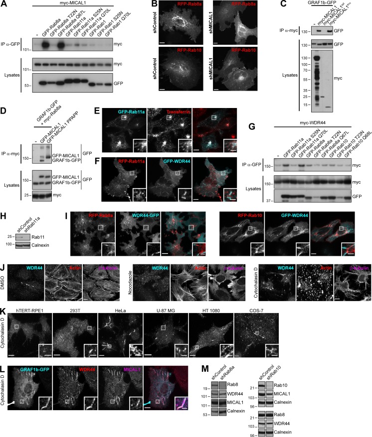
Figure S4.
MICAL1 and WDR44 connect GRAF1b/2 to Rab8, Rab10, and Rab11. (A, C, D, and G) Immunoprecipitation (IP) of transfected 293T cells with α-GFP (A and G), α-myc–coated beads (C), or α-myc (D). (A) myc-MICAL1 was coimmunoprecipitated by Rab8a Q67L, but not by Rab8a T22N. (B) Confocal stacks of shControl or shMICAL1-expressing HeLa cells transfected with RFP-Rab8a or RFP-Rab10. (C) myc-MICAL1 and myc-MICAL1Pro but not myc-MICAL1tail coimmunoprecipitated GRAF1b-GFP. (D) GRAF1b-GFP was only coimmunoprecipitated with myc-Rab8a when GFP-MICAL1 was coexpressed, not GFP–MICAL1 PPAPP. (E) Confocal images of GFP-Rab11a–transfected HeLa cells incubated with Alexa Fluor 546–Transferrin (10 µg/ml, 30 min) showing colocalization. (F) Confocal images of transfected HeLa cells. RFP-Rab11a was found on GFP-WDR44–positive tubules, but GFP-WDR44 was not recruited to RFP-Rab11a–positive endosomes. (G) myc-WDR44 was coimmunoprecipitated by Rab11a and by its constitutively active mutant Rab11a Q70L, but not by the dominant negative mutant Rab11a S25N. (H and M) Western blot analysis of cell lysates from HeLa cells transfected with specific shRNA-encoding plasmids. Calnexin was used as loading control. (H) Rab11 was knocked down in shRab11a-transfected cells. (I) Confocal images of transfected HeLa cells showing RFP-tagged Rab8a/10 on the same intracellular tubules as GFP-tagged WDR44 but enriched on complementary segments of the tubules. (J) Confocal stacks of HeLa cells incubated with DMSO (vehicle, 2 h), Nocodazole (20 µg/ml, 2 h), or Cytochalasin D (0.5 µg/ml, 30 min) and stained with α-WDR44, α-β-tubulin, and Alexa Fluor 546–phalloidin. (K) Confocal stacks of hTERT-RPE1, 293T, HeLa, U-87 MG, HT 1080, and COS-7 cells incubated with Cytochalasin D (0.5 µg/ml, 30 min) and stained with α-WDR44. Cytochalasin D induced endogenous WDR44 tubules in HeLa, HT 1080, and COS-7 cells (compare with Fig. S3 E). (L) Confocal images of transfected HeLa cells incubated with Cytochalasin D (0.5 µg/ml, 30 min) and stained with α-WDR44 and α-MICAL1 showing colocalization with GRAF1b-GFP. (M) Rab8a and Rab10 were knocked down in shRab8a- and shRab10-transfected cells, respectively. In each case, the two parts of the blots are from the same membrane and correspond to identical exposure times. shRab10: the upper three blots and the lower three are from replicate membranes. (B, E, F, I, and J–L) Insets show magnifications of the boxed areas. Scale bars: 10 µm; scale bars of insets: 2 µm.

